
Doppler in Critical Care: Hemodynamic Monitoring and Beyond
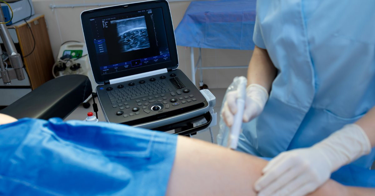
A blood pressure reading can signal danger without revealing the cause. Critical care teams still need to know whether the heart pumps effectively, whether blood reaches vital organs, and whether an intervention improves circulation. Doppler ultrasound expands hemodynamic monitoring by converting moving blood into audible signals, waveforms, and color flow.
That information can sharpen clinical reasoning in shock, respiratory failure, neurologic emergencies, and postoperative deterioration. Doppler also supports repeated assessment, providing clinicians with a dynamic view rather than a single snapshot. Trained professionals should correlate each finding with the physical examination, laboratory data, and the patient’s response over time.
A Faster View of Flow
Doppler technology detects shifts in the frequency of reflected ultrasound as red blood cells move. The magnitude of the shift relates to blood velocity, while the direction indicates whether blood is moving toward or away from the transducer. Pulsed-wave Doppler samples flow at a selected location, continuous-wave Doppler captures higher velocities along the beam, and color Doppler maps direction and average velocity across the image.
Each mode addresses a distinct clinical question. A handheld audio unit can help a clinician locate a peripheral arterial signal, whereas echocardiography can measure intracardiac flow and relate it to anatomy. Clinicians should match the device, probe, and mode to the intended assessment rather than treating Doppler as a single interchangeable technology.
Hemodynamics at the Bedside
Cardiac Doppler adds quantitative flow data to a focused cardiac examination. Clinicians can measure the velocity-time integral in the left ventricular outflow tract and, together with the outflow tract area, estimate stroke volume. Multiplying stroke volume by heart rate yields an estimate of cardiac output. Serial measurements can show whether output rises, falls, or remains unchanged after an intervention.
Doppler also helps teams assess more than forward flow. Transmitral and tissue Doppler patterns can provide information about left-sided filling pressures when the clinical setting supports those measurements. Continuous-wave Doppler can capture high velocities across valves, while right-heart measurements can reveal pressure overload and impaired right ventricular performance. Clinicians should interpret several findings together rather than relying on a single number.
Tracking Treatment Response
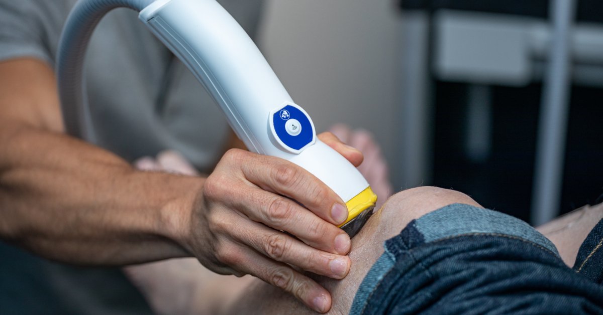
Critical care rarely stands still, so hemodynamic assessment shouldn’t end after a single examination. A team can repeat the same Doppler measurement before and after a fluid challenge, passive leg raise, diuretic dose, inotrope adjustment, or ventilator change. A consistent acquisition method enables the team to compare trends with greater confidence.
Echocardiography can track right- and left-sided hemodynamics over minutes, hours, or days, but it doesn’t provide continuous monitoring. The clinician must return to the same view, carefully align the beam, and place the sample volume at the same location. The chart should record the intervention, heart rhythm, ventilator settings, blood pressure, and acquisition time.
Reading Shock Patterns
Doppler can help differentiate physiologic problems that produce similar vital signs. Low forward flow may indicate poor ventricular contraction, inadequate filling, severe valvular obstruction, or increased right ventricular afterload. Abnormal venous and intracardiac flow patterns may also support concern for congestion or elevated filling pressure.
No Doppler value alone identifies the cause of shock. A clinician should integrate flow data with chamber size, ventricular motion, lung findings, pericardial assessment, vascular examination, laboratory values, and perfusion findings. That approach helps teams distinguish a patient who may respond to increased preload from one whose circulation could worsen with additional volume.
Peripheral Circulation
Handheld medical Dopplers extend bedside assessment beyond the heart. Clinicians can use an appropriate vascular probe to detect arterial or venous flow, assess difficult-to-palpate peripheral signals, and support ankle-brachial index measurement when cuff pressures and peripheral signals are technically reliable. Available devices range from audio-only models to systems that display direction or waveforms.
A focused peripheral check can help teams monitor distal perfusion after arterial catheter placement, vascular procedures, trauma, or sudden limb changes. A returning signal can support the examination, while a weak or absent signal can prompt escalation per local protocols. A handheld unit doesn’t replace duplex imaging, angiography, or specialist evaluation when clinicians suspect acute limb ischemia.
Cerebral Monitoring
Transcranial Doppler brings flow assessment to the brain. Neurocritical care teams can track blood velocity in major intracranial arteries, monitor evolving vasospasm after subarachnoid hemorrhage, and assess changes in cerebral perfusion patterns. The bedside format is particularly valuable when a neurologically unstable patient can’t safely travel to a scanner.
The technique requires specialized training, consistent insonation windows, and careful interpretation. Skull anatomy can limit the acoustic window, and systemic blood pressure, carbon dioxide, heart rate, and vessel anatomy can influence velocity. Transcranial Doppler complements computed tomography, magnetic resonance imaging, angiography, neurologic examination, and invasive monitoring rather than carrying the full diagnostic burden.
Procedures and Device Support
Color Doppler can help clinicians distinguish arteries from veins, confirm flow direction, and assess vessel patency during procedure planning. Point-of-care ultrasound programs also use ultrasound for resuscitation, diagnosis, physiologic monitoring, and procedural guidance. Echocardiographic Doppler can also support ventricular assist device adjustment and weaning protocols.
The exact role depends on the equipment and the clinician’s credentials. An audio-only vascular Doppler can detect a signal but can’t visualize surrounding anatomy. An imaging system can display vessel depth, neighboring structures, and flow direction, and it supports assessment for thrombosis or hematoma. Clear role definitions prevent teams from asking a device to answer a question outside its intended use.
Technique Shapes the Result
Doppler accuracy depends on beam alignment, probe frequency, sample placement, gain, scale, and patient physiology. Poor beam alignment can underestimate velocity. Excessive gain can introduce noise, an incorrect scale can hide or distort flow, and inconsistent placement can make serial comparisons unreliable. Arrhythmias also require representative beats rather than a single convenient cycle.
Mechanical ventilation alters intrathoracic pressure, vasoactive drugs modify vascular tone, dressings restrict access, and pain or agitation can limit positioning. Teams should document technical limitations rather than force certainty from weak data.
Building a Reliable Workflow
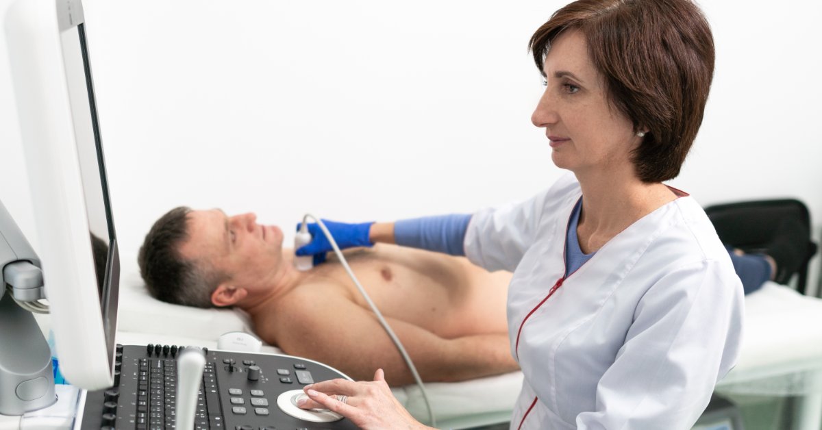
A reliable Doppler program begins with a clear clinical question and a standardized acquisition process. Teams need competency standards, cleaning procedures, documentation rules, maintenance plans, and ready access to the appropriate probes, gel, batteries, and charging equipment. Quality review should cover signal acquisition, interpretation, and clinical integration.
Cascade Health Care serves healthcare professionals and organizations across hospital, emergency response, community health, and educational settings. That audience reflects the environments where Doppler skills can support patient assessment. Equipment selection should follow the device’s intended use, the users’ training, and the decisions the team needs to make.
Flow That Informs Action
Doppler provides critical care teams with hemodynamic monitoring and beyond. It shows how blood moves, how the heart responds, how peripheral perfusion changes, and how cerebral flow evolves. The strongest interpretation comes from repeated, technically sound measurements that answer a specific question.
When clinicians integrate Doppler findings with anatomy, examination, and clinical trajectory, the technology becomes more than a signal detector. It becomes a practical way to test a hemodynamic hypothesis, assess response, and recognize when the patient needs formal imaging or specialist assessment. In a setting where physiology can change within minutes, that clearer view of flow can support faster, more disciplined decisions.
Recent Posts
-

Pros and Cons of Custom Birth Kits vs. Pre-Assembled Kits
Preparing for a birth means thinking beyond individual products. Healthcare professionals need a rel
-

How To Choose the Best Fetal Doppler for Midwifery Practice
Choosing a fetal Doppler can feel straightforward until you start comparing models. Midwives need eq