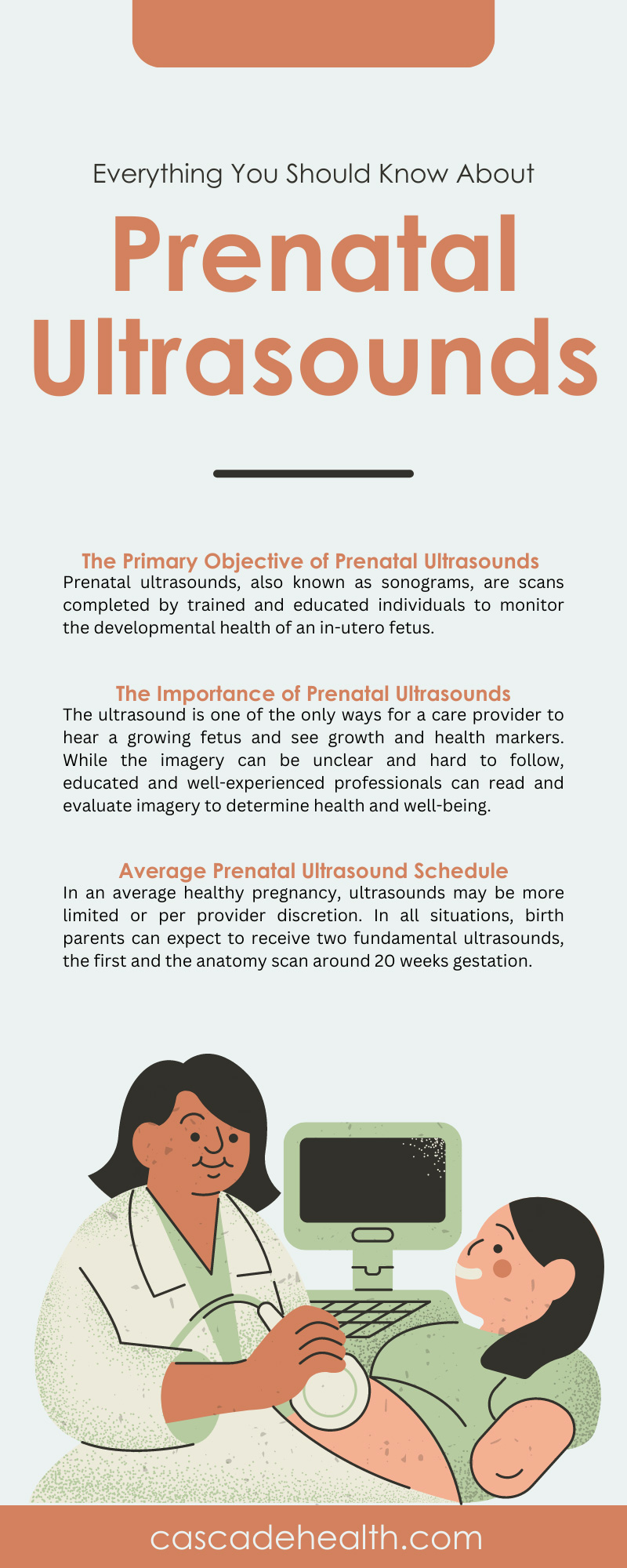
Everything You Should Know About Prenatal Ultrasounds
One of the most important elements of routine prenatal care and guidance is receiving or administering a prenatal ultrasound. These vital and special scans allow providers the opportunity to explore the overall health and viability of a growing fetus, in addition to gaining information about position, size, and sex.
Expecting parents look forward to each ultrasound as it’s a chance to connect with their new baby as they await the official meet and greet. And some birth parents agree that seeing those small but mighty features on the screen leads to love at first sight. There is a lot to know about prenatal ultrasounds, so we gathered everything worth knowing for you here.
The Primary Objective of Prenatal Ultrasounds
Prenatal ultrasounds, also known as sonograms, are scans completed by trained and educated individuals to monitor the developmental health of an in-utero fetus. Ultrasound technicians, also known as sonographers, certified nurse midwives, and obstetricians, can complete ultrasounds and read the scans once complete.
The two most common reasons for an ultrasound are developmental monitoring and assessing the fetus if a problem presents itself. During developmental monitoring, the primary objective is to measure healthy growth markers. Should a provider suspect an issue, whether mother or fetus-related, they may complete an ultrasound to rule out various contributing factors, if any apply.
An ultrasound machine uses distinct sound wave technology to bounce off the interior bodily structures and capture sounds and images. These sound waves take the captured data and report back onto a screen what is detectable both visually and audibly. In general, prenatal ultrasounds are safe and ethical. But care providers should only administer them when medically necessary according to the birth parent’s gestational experience.
The Importance of Prenatal Ultrasounds
The ultrasound is one of the only ways for a care provider to hear a growing fetus and see growth and health markers. While the imagery can be unclear and hard to follow, educated and well-experienced professionals can read and evaluate imagery to determine health and well-being. It’s important for birth parents to understand how well their growing fetus is developing and remain aware of any health trends should they become detectable before labor and delivery.
What Can We Learn From Prenatal Ultrasounds?
We mentioned prenatal ultrasounds being valuable in developmental health monitoring and risk evaluation, but providers can also learn various things that outline one or both of these circumstances. In the average pregnancy, ultrasounds remain a positive experience where birth parents get to hear and see their baby and leave with a few images. But there are times when these experiences do not go as planned, and a provider may learn about issues surrounding the health and well-being of either the mother or the baby.
Providers generally use ultrasound to confirm the pregnancy, determine the type of pregnancy, whether ectopic, molar, or miscarriage, declare gestation age and approximate due date, heart rate, and movements, confirm the number of fetal heartbeats, and complete evaluation of the mother’s amniotic fluid levels and ovarian health. In almost all cases, a provider can learn about issues with the baby’s developing organs, bones, or muscles and understand the position. The position plays a role in labor and delivery, so it’s essential to understand this element.
With everything there is to know about prenatal ultrasounds, it’s essential to understand that there are two types that providers will rely on. Each one serves a fundamental purpose, and providers will use the scanning method that medically suits the patient, fetus, and objectives for the scan.
Transvaginal Ultrasounds
A transvaginal ultrasound is an interior-positioned scanning method. The provider will insert a probe-like device into the vaginal canal and collect the same data as an abdominal ultrasound. Providers rely on transvaginal ultrasounds during early pregnancy to gain more clarity when the fetus is incredibly small. From here, they can detect the fetal heartbeat, declare the estimated due date through gestational age, and receive clearer imagery compared to the abdominal ultrasound. As the pregnancy evolves and the fetus grows, an abdominal ultrasound will suffice.
Abdominal Ultrasounds
The abdominal ultrasound gets completed externally, with a transducer, gel, and a relay screen where the images go. The applied gel helps the transducer receive more accurate data and helps move the transducer around the abdominal area easily. Because the transvaginal ultrasound helps provide better images and sounds in early pregnancy, a provider will not rely on an abdominal scan before 12 weeks of gestation.
Average Prenatal Ultrasound Schedule
Because prenatal ultrasounds should only get used when medically appropriate and necessary, the scan schedule is minimal and often varies case by case. If a provider deems a pregnancy high-risk, if the birth mother develops a health condition, or if the provider suspects issues with the growing fetus, ultrasounds may occur more frequently. In an average healthy pregnancy, ultrasounds may be more limited or per provider discretion. In all situations, birth parents can expect to receive two fundamental ultrasounds, the first and the anatomy scan around 20 weeks gestation.
The Importance of the First Ultrasound
The first ultrasound is special and outlines the beginning of a new chapter in the birth parents’ lives. This is the confirmation ultrasound that declares a patient is carrying a pregnancy at this time and also helps date the expected arrival of this new bundle of joy. The first ultrasound can happen as early as seven weeks gestation but will be a transvaginal scan for accuracy. Depending on the provider, they may request to wait until 12 weeks of estimated gestation to perform the first ultrasound.
The Anatomy Scan
The second most important milestone and completed scan patients can expect is the anatomy scan between 18 and 20 weeks gestation. This ultrasound is important because the fetus is much larger, and more data is available. In this scan, providers can learn about the sex of the fetus and potentially gain insight into possible congenital disabilities. The brain, bones, and heart are all still developing, but issues are more detectable at this time if any present themselves.
Cascade Health Care values the importance and integrity of safe and ethical prenatal ultrasounds. We support all medical professionals and organizations in their quest to achieve high-quality maternity care and offer trusted ultrasounds with a pregnancy doppler safe for birth parents and babies. Shop our entire collection of dopplers and supplies today to provide the best care to each family.

Recent Posts
-
The History and Evolution of the Speculum
Most women view the speculum as a symbol of modern routine healthcare, a cold but necessary instrume
-
What Midwives Should Teach New Parents Regarding Breastmilk
The transition into parenthood presents a profound physiological and emotional shift for growing fam


