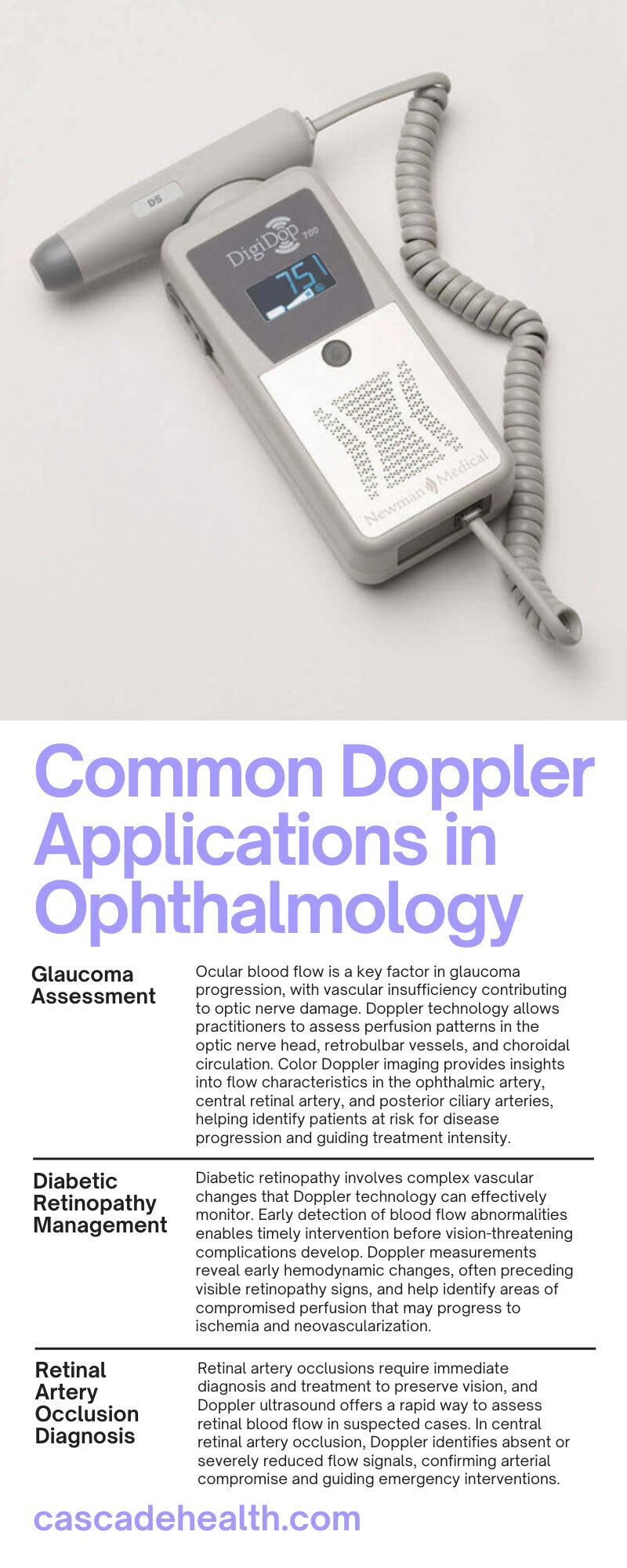
6 Common Doppler Applications in Ophthalmology
Doppler ultrasound technology has revolutionized diagnostic capabilities across multiple medical specialties, with ophthalmology standing as one of the most innovative adopters. Eye care professionals now rely on Doppler assessments to evaluate blood flow patterns, detect vascular abnormalities, and monitor treatment responses with unprecedented precision.
This comprehensive guide explores six common Doppler applications in ophthalmology. From glaucoma assessment to tumor evaluation, these diagnostic techniques provide critical insights that enhance patient outcomes and treatment decisions.
Understanding Doppler Technology
Doppler ultrasound operates on the principle of frequency shift detection when sound waves encounter moving objects. The technology measures changes in frequency as ultrasound waves reflect off moving blood cells, creating detailed information about blood flow velocity, direction, and volume.
Advanced Doppler systems combine multiple imaging modes to provide comprehensive vascular assessments. Color Doppler mapping overlays flow information onto grayscale images, while spectral Doppler analysis quantifies specific flow parameters. Power Doppler enhances sensitivity to low-velocity flow, making it particularly valuable for detecting subtle perfusion changes in ocular tissues.
Modern ophthalmic Doppler units feature specialized high-frequency transducers optimized for superficial tissue penetration. These devices deliver exceptional resolution for evaluating delicate ocular structures while maintaining patient comfort during examinations.
Why Dopplers Are Essential in Ophthalmology
Vascular health directly impacts visual function, making blood flow assessment crucial for comprehensive eye care. The eye's complex vascular network includes retinal arteries, choroidal circulation, and optic nerve perfusion—each playing vital roles in maintaining vision.
Doppler technology enables non-invasive evaluation of these critical blood flow patterns, allowing practitioners to detect early pathological changes before irreversible damage occurs. This early detection capability transforms treatment approaches from reactive to preventive, significantly improving long-term visual outcomes.
Clinical decision-making benefits tremendously from objective flow measurements that Doppler technology provides. Rather than relying solely on subjective observations, practitioners access quantifiable data that supports evidence-based treatment protocols and monitoring strategies.
Common Ophthalmology Doppler Applications
Six primary applications demonstrate Doppler ultrasound's versatility in ophthalmic practice. These applications span diagnostic, monitoring, and prognostic functions across multiple ocular conditions. Each application leverages specific Doppler capabilities to address unique clinical challenges in eye care.
Glaucoma Assessment
Ocular blood flow is a key factor in glaucoma progression, with vascular insufficiency contributing to optic nerve damage. Doppler technology allows practitioners to assess perfusion patterns in the optic nerve head, retrobulbar vessels, and choroidal circulation. Color Doppler imaging provides insights into flow characteristics in the ophthalmic artery, central retinal artery, and posterior ciliary arteries, helping identify patients at risk for disease progression and guiding treatment intensity.
Spectral Doppler analysis quantifies resistance indices and peak systolic velocities in retrobulbar vessels, with elevated resistance patterns often correlating with disease severity and treatment response. Monitoring flow parameters alongside intraocular pressure measurements offers a more comprehensive approach to glaucoma management, complementing traditional techniques with additional prognostic information.
Doppler evaluation is beneficial in normal-tension glaucoma, where vascular factors play a larger role than pressure-related damage. These assessments help identify patients who may benefit from vascular-protective therapies in addition to conventional pressure-lowering treatments, enhancing personalized care strategies.
Diabetic Retinopathy Management
Diabetic retinopathy involves complex vascular changes that Doppler technology can effectively monitor. Early detection of blood flow abnormalities enables timely intervention before vision-threatening complications develop. Doppler measurements reveal early hemodynamic changes, often preceding visible retinopathy signs, and help identify areas of compromised perfusion that may progress to ischemia and neovascularization.
Color Doppler mapping highlights neovascular flow patterns associated with proliferative diabetic retinopathy, helping practitioners distinguish active neovascularization from inactive fibrovascular tissue. This information guides treatment decisions, such as when to use anti-VEGF therapy or laser photocoagulation.
Choroidal blood flow evaluation offers valuable insights into diabetic macular edema pathogenesis and treatment responses. Doppler measurements correlate with macular thickness and visual acuity changes, helping optimize anti-VEGF injection protocols and monitor treatment efficacy.
Retinal Artery Occlusion Diagnosis
Retinal artery occlusions require immediate diagnosis and treatment to preserve vision, and Doppler ultrasound offers a rapid way to assess retinal blood flow in suspected cases. In central retinal artery occlusion, Doppler identifies absent or severely reduced flow signals, confirming arterial compromise and guiding emergency interventions.
For branch retinal artery occlusions, Doppler precisely localizes flow abnormalities, helping determine the extent of the occlusion and predict visual field defects. This targeted assessment supports tailored treatment and informed discussions with patients about prognosis.
Doppler evaluation also distinguishes arterial occlusions from other causes of sudden vision loss, such as optic neuropathies or retinal detachments. This ensures accurate diagnosis, prevents inappropriate treatments, and optimizes emergency patient care.
Choroidal Neovascularization Detection
Age-related macular degeneration and other conditions create choroidal neovascular membranes that Doppler technology can effectively identify. Early detection with Doppler enables timely anti-VEGF treatments, helping preserve central vision.
Power Doppler imaging is highly sensitive to low-velocity blood flow within choroidal neovascular complexes, often detecting subtle flow signals before angiographic evidence appears. This makes it especially useful for identifying occult membranes and enabling earlier intervention.
Color Doppler helps differentiate classic from occult neovascularization based on flow patterns. These distinctions guide the selection of anti-VEGF agents and treatment protocols, with Doppler also aiding in monitoring therapy effectiveness. Reduced flow signals after treatment often correlate with improved anatomy and stabilized vision.
Ocular Tumor Evaluation
Intraocular tumors exhibit distinct vascular patterns detectable via Doppler ultrasonography, aiding in the diagnosis and treatment of uveal melanomas, metastases, and benign lesions. Malignant tumors, such as melanomas, display high-velocity, low-resistance flow patterns, while benign lesions typically show minimal vascular activity, complementing morphological features in diagnostic workflows.
Doppler assessments play a key role in predicting metastatic risk and guiding treatment intensity. Highly vascular melanomas are associated with greater metastatic potential, influencing surveillance strategies and adjuvant therapy decisions. This vascular data provides valuable prognostic insights beyond traditional tumor measurements.
Serial Doppler evaluations help monitor treatment response after radiation therapy or surgery. Decreased vascular activity signals effective treatment, while persistent blood flow may indicate residual tumor activity, prompting further intervention.
Where To Find the Best Dopplers
At Cascade Health Care, we provide medical practitioners with reliable, high-quality vascular Dopplers from top manufacturers like Newman, Huntleigh, Imex, and Wallach/Summit. Our goal is to ensure you have access to the most advanced Doppler technology for accurate diagnostics.
We’re proud to offer competitive pricing, multi-unit discounts, and zero sales tax on all our vascular Dopplers for sale, making cutting-edge technology accessible to practices of all sizes. Whether you’re a solo practitioner or part of a large medical center, we have solutions tailored to your needs.
Our expert customer service team is here to guide you in selecting the right Doppler system for your specific clinical applications. Explore our complete selection today to find the perfect match for your diagnostic requirements.
Advancing Ophthalmic Care Through Vascular Assessment
Doppler ultrasound technology has fundamentally transformed ophthalmology by providing non-invasive access to critical vascular information. These six Doppler applications in ophthalmology represent just the beginning of Doppler's potential in eye care, with emerging techniques continuously expanding diagnostic possibilities.
Investment in quality Doppler equipment represents a commitment to excellence in patient care and practice growth. The diagnostic insights these tools provide create lasting value for practitioners and patients, establishing foundations for superior clinical outcomes and practice success.

Recent Posts
-
Best Practices for Cleaning and Maintaining Pulse Oximeters
Midwives and medical professionals understand the critical nature of accurate patient data. When mon
-
What To Do With Birthing Supplies After a Home Birth
Home birth is on the rise. As families increasingly seek the comfort and autonomy of birthing in the


