5 Medical Instruments Used During IUD Procedures
Intrauterine device insertion requires proper technique and the right medical instruments to ensure patient safety and procedural success. Medical professionals who perform these procedures rely on specialized tools designed specifically for gynecological examinations and IUD placements. Understanding the function and proper use of each instrument enhances procedural efficiency and patient comfort.
This comprehensive guide explores five medical instruments used during IUD procedures, providing medical professionals with essential knowledge about the equipment that supports safe and effective contraceptive care. Each instrument serves a distinct purpose in the insertion process, from initial examination to final placement verification.
1. Vaginal Speculum
The vaginal speculum serves as the foundation for IUD insertion by providing clear visualization of the cervix throughout the procedure. Medical professionals select from various speculum types, including Graves, Pedersen, and disposable models, based on patient anatomy and comfort considerations. The instrument holds the vaginal walls apart, creating an unobstructed view of the cervical os and surrounding tissue.
Proper speculum placement minimizes patient discomfort while maximizing visibility for accurate instrument guidance. Many practices maintain a supply of speculums that includes multiple sizes to accommodate diverse patient needs. Modern speculums feature smooth edges, ergonomic designs, and warming capabilities that enhance the patient experience during an examination.
The choice between metal and plastic options depends on facility sterilization protocols, cost considerations, and environmental preferences. Medical professionals must warm metal speculums before insertion to prevent patient discomfort from cold instruments. Adequate lighting, combined with proper speculum positioning, enables clinicians to identify the anatomical landmarks necessary for safe IUD placement.
2. Tenaculum

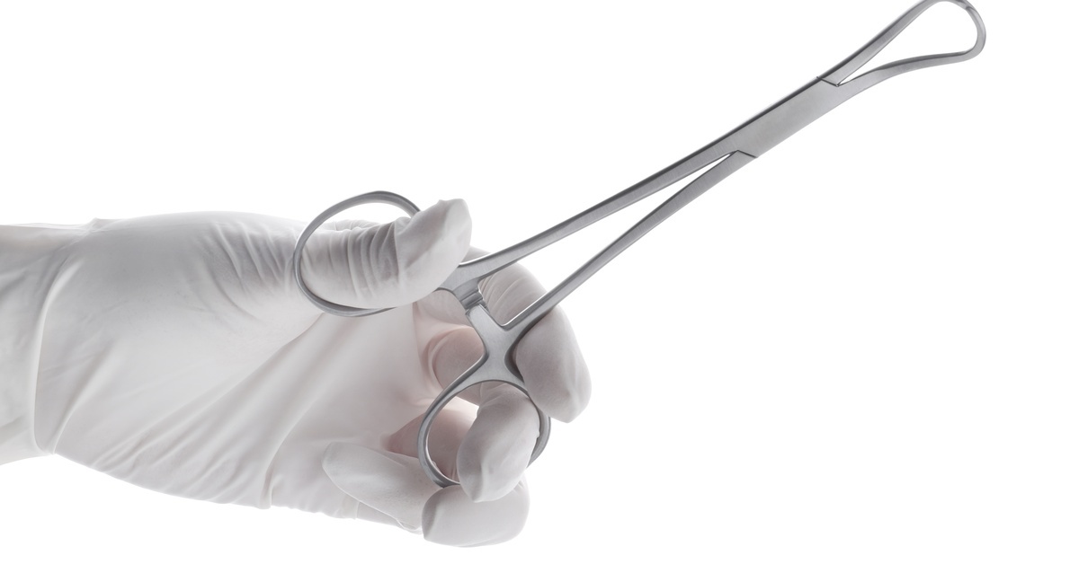
The tenaculum stabilizes the cervix during IUD insertion, providing the traction necessary for proper uterine alignment and instrument passage. This grasping instrument features sharp teeth or hooks that secure the anterior cervical lip, straightening the cervical canal and uterine axis.
Single-tooth and double-tooth variations offer different levels of grip strength depending on cervical tissue characteristics and procedural requirements. The tenaculum reduces cervical movement during sound measurement and IUD insertion, thereby improving procedural accuracy and decreasing perforation risk.
Medical professionals apply the tenaculum gently but firmly to maintain cervical position without causing excessive tissue trauma. Some clinicians prefer using a local anesthetic at the tenaculum application site to minimize patient discomfort during the procedure.
The instrument's design allows medical professionals to manipulate control of a patient’s cervical position, which proves especially valuable when encountering a severely anteverted or retroverted uterus. After IUD placement, clinicians carefully remove the tenaculum and assess the application site for bleeding, applying pressure if necessary to achieve hemostasis.
3. Uterine Sound
The uterine sound measures the depth and orientation of the uterine cavity before IUD insertion, providing critical information to prevent improper placement and complications. This slender, graduated instrument allows clinicians to determine the exact length of the uterine cavity, which guides proper IUD positioning in the fundal region.
The sound helps identify uterine position, cavity abnormalities, and potential obstacles that might complicate insertion. Medical professionals advance the sound gently through the cervical canal until reaching the uterine fundus, noting the measurement at the external cervical os.
The information gained from sounding directly influences IUD placement depth and reduces the risk of expulsion or perforation. Key measurements obtained during sounding include:
- Uterine cavity depth (typically 6-9 centimeters in adult women)
- Cervical canal length and any stenosis present
- Uterine axis orientation (anteversion, retroversion, or midposition)
- Presence of fibroids, polyps, or structural abnormalities
4. IUD Insertion Tube
The IUD insertion tube delivers the contraceptive device through the cervical canal into the uterine cavity with precision and minimal trauma. This sterile, single-use instrument contains the folded IUD within its barrel and features measurement markings that correspond to uterine cavity depth.
The tube’s design protects the IUD from contamination during passage through the vaginal and cervical environments. Medical professionals load the IUD into the insertion tube according to the manufacturer’s specifications, ensuring proper device orientation before beginning the insertion process.
The tube’s diameter remains small enough to pass through the cervical canal while maintaining sufficient rigidity for controlled advancement. Clinicians advance the insertion tube to the predetermined depth based on prior uterine sounding measurements, typically positioning the IUD in the upper uterine cavity near the fundus.
The plunger mechanism releases the IUD arms at the appropriate depth, allowing the device to expand within the uterine cavity. Steadily withdrawing the insertion tube after IUD deployment prevents device displacement and ensures proper positioning.
5. Curved Scissors

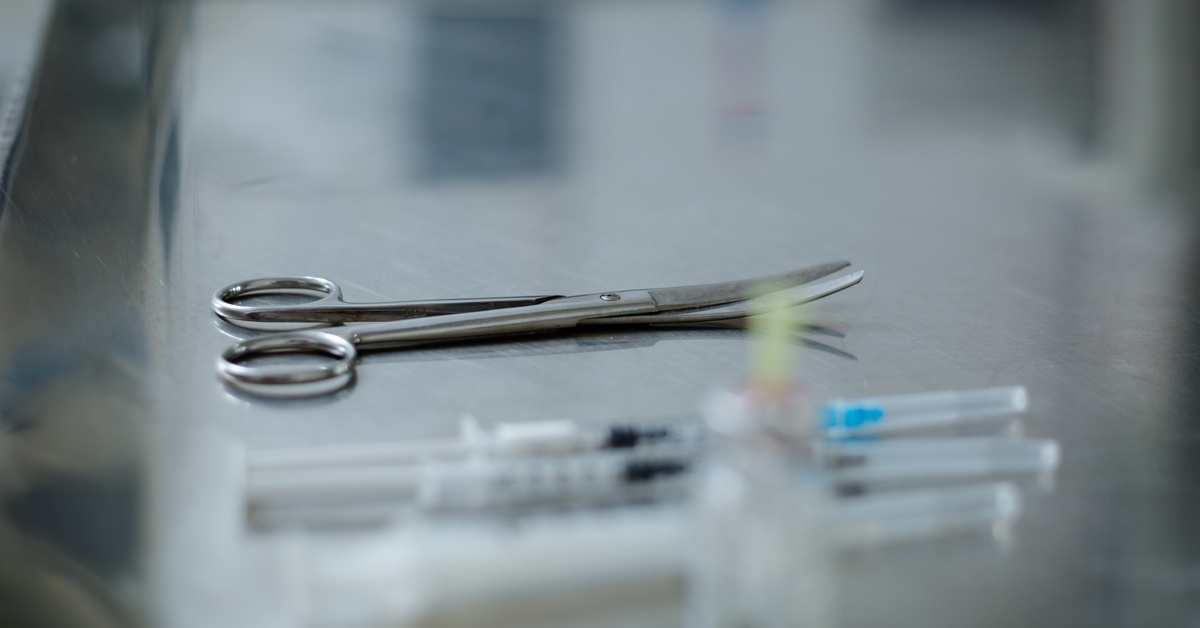
Curved scissors complete the IUD insertion procedure by trimming the device strings to the appropriate length for patient comfort and future removal. These specialized surgical scissors feature curved blades that provide excellent visualization during the cutting process within the vaginal canal.
Medical professionals trim IUD strings to extend approximately three to four centimeters beyond the external cervical os, balancing patient comfort with accessibility for future removal. The curved design allows clinicians to maintain a clear view of the cutting point while maneuvering around the speculum and cervical anatomy.
Proper string length prevents the strings from protruding through the cervical os in a way that might cause partner awareness during intercourse or patient discomfort. Excessively long strings can irritate the vaginal walls or become visible outside the vagina, while strings trimmed too short can complicate removal procedures.
The scissors must remain sharp to achieve clean cuts that prevent string fraying, which could cause irritation or infection. After trimming, clinicians verify string placement and document the final string length in the patient's medical record for future reference.
Support Procedural Success With the Proper Tools
Successful IUD insertion depends on medical professionals having access to quality instruments designed specifically for these procedures. These five medical instruments used during IUD procedures work together to ensure safe, efficient, and comfortable contraceptive placement.
Each instrument plays a key role throughout the procedure, from visualizing the cervix to trimming the IUD strings. Medical professionals who understand the purpose and proper use of each tool can optimize procedural outcomes while minimizing patient discomfort and complication risks.
Cascade Health Care provides medical professionals with reliable access to high-quality instruments necessary for gynecological procedures, supporting excellence in patient care. Investing in the appropriate equipment and maintaining up-to-date knowledge of instrument selection will enhance your clinical practice and demonstrate a commitment to patient safety and procedural success.
Regularly inspecting and maintaining all IUD instruments ensures longevity, precise performance, and patient safety, reducing procedural complications and maintaining consistent clinical outcomes. Providing staff with ongoing training in instrument handling and placement techniques further enhances procedural efficiency, patient comfort, and confidence during IUD insertions. Clinics looking to expand their inventory will find the proper speculum for sale at Cascade Health Care, designed to meet every patient’s needs.
Recent Posts
-
Best Practices for Cleaning and Maintaining Pulse Oximeters
Midwives and medical professionals understand the critical nature of accurate patient data. When mon
-
What To Do With Birthing Supplies After a Home Birth
Home birth is on the rise. As families increasingly seek the comfort and autonomy of birthing in the


